
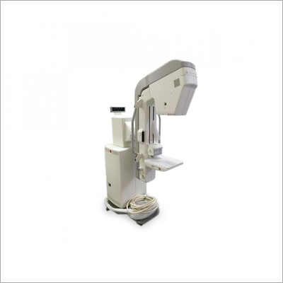
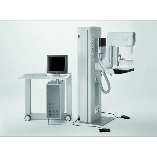
GE Senographe DMR Mammography
Product Details:
- Properties Efficient, Reliable, High-Resolution Imaging
- Frequency 50/60 Hz
- Product Type Digital Mammography System
- Weight Approx. 380 kg
- Dimension (L*W*H) Approx. 1900 mm x 800 mm x 2100 mm
- Material High-quality Composite and Metal Alloys
- Display Type Digital Display
- Click to View more
X
GE Senographe DMR Mammography Price And Quantity
- 10
- High-sensitivity digital flat panel detector
- Image enhancement, annotation, storage, print
- High (up to 20 lp/mm)
- 24 x 30 cm
- Touchscreen operation with menu navigation
- Padded compression paddle, ergonomic design
- Automatic and manual compression available
- Senographe DMR
- 18 x 24 cm, 24 x 30 cm
- Option for 3D breast tomosynthesis upgrade
- Floor-mounted gantry
- LED collimator for precise positioning
- 2D, Spot Compression View, Magnification View
- Up to 10 patients per hour (depending on workflow)
- Standard and Magnification Views
- DICOM compatible network connectivity
- Emergency stop, mechanical interlock, dose monitoring
- 0.1/0.3 mm
- Molybdenum/Rhodium target, dual filter
- Removable anti-scatter grid
GE Senographe DMR Mammography Product Specifications
- +15C to +32C Operation
- Adjustable: 1200 1800 mm
- Full-field mammography
- Ergonomic, Mobile Gantry Design
- 220 240 V AC
- High accuracy for microcalcification detection
- Digital image storage
- Single Phase
- Breast Imaging Examination
- Mammographic Screening and Diagnosis
- Semi-automatic
- Digital LCD Controller
- Hospitals, Diagnostic Centers, Clinics
- 30% 75% non-condensing
- 2.0 kVA
- Non-invasive Diagnostic Imaging
- Standard Medical White with Grey Accents
- Digital Display
- High-quality Composite and Metal Alloys
- Approx. 380 kg
- Electric
- Approx. 1900 mm x 800 mm x 2100 mm
- Efficient, Reliable, High-Resolution Imaging
- Digital Mammography System
- 50/60 Hz
- High-sensitivity digital flat panel detector
- Image enhancement, annotation, storage, print
- High (up to 20 lp/mm)
- 24 x 30 cm
- Touchscreen operation with menu navigation
- Padded compression paddle, ergonomic design
- Automatic and manual compression available
- Senographe DMR
- 18 x 24 cm, 24 x 30 cm
- Option for 3D breast tomosynthesis upgrade
- Floor-mounted gantry
- LED collimator for precise positioning
- 2D, Spot Compression View, Magnification View
- Up to 10 patients per hour (depending on workflow)
- Standard and Magnification Views
- DICOM compatible network connectivity
- Emergency stop, mechanical interlock, dose monitoring
- 0.1/0.3 mm
- Molybdenum/Rhodium target, dual filter
- Removable anti-scatter grid
GE Senographe DMR Mammography Trade Information
- 10 Per Month
- 1 Months
Product Description
Specification
FDA Clearance Yes
Focal Spots Large 0.3 mm
Focal Spots Small 0.1 mm
High Frequency Generator 5 kW
mAs Range 4-500 mAs
Source to Image Distance (SID) 66 cm
Type Digital
Equipment Type Mammogram Machine
X-ray release options Hand switch ,Control console
Brand GE Healthcare
Advanced Imaging Precision
With a high-sensitivity digital flat panel detector and a resolution of up to 20 lp/mm, the GE Senographe DMR offers unmatched clarity for detecting even the smallest breast abnormalities. Its dual-filter X-ray tube and removable anti-scatter grid ensure detailed, high-quality mammographic images for confident diagnosis.
Optimized Workflow and Patient Comfort
Designed for busy clinical environments, this mammography system needs only a single phase, allowing up to 10 patients per hour depending on workflow. The ergonomic gantry allows adjustable height, a padded compression paddle reduces discomfort, and the intuitive touchscreen panel supports efficient operation.
Future-Ready and Reliable
Equipped with digital storage, DICOM compatibility, and software for image enhancement, the GE Senographe DMR supports easy data management. The system can be upgraded to 3D breast tomosynthesis for enhanced diagnostic capability, ensuring long-term value for medical imaging providers.
FAQs of GE Senographe DMR Mammography:
Q: How does the GE Senographe DMR improve mammographic screening efficiency?
A: The Senographe DMR streamlines workflow with a digital flat panel detector and semi-automatic operation, enabling technicians to image up to 10 patients per hour. Its touchscreen interface and fast image processing minimize delays and support efficient screening in high-volume settings.Q: What are the key benefits of the high-sensitivity digital detector in this system?
A: The high-sensitivity flat panel detector provides high-resolution images up to 20 lp/mm, facilitating the reliable detection of microcalcifications and subtle tissue changes. This leads to more accurate diagnoses, especially in early breast cancer detection.Q: When should magnification and spot compression views be used?
A: Magnification and spot compression views are employed when detailed evaluation of suspicious areas or microcalcifications is required. These imaging modes enhance visualization, allowing radiologists to assess small lesions and subtle abnormalities with higher precision.Q: Where can the GE Senographe DMR be installed?
A: This system is suitable for installation in hospitals, diagnostic centers, and clinics. Its floor-mounted, ergonomic design fits standard imaging suites and meets the needs of both screening programs and diagnostic assessments in diverse healthcare environments.Q: What process ensures patient safety during mammography on this system?
A: Patient safety is prioritized through built-in features like emergency stop, mechanical interlock, and dose monitoring. The system uses padded compression paddles and precise LED collimation to optimize comfort while maintaining accurate, low-dose imaging protocols.Q: How is image data managed and stored with this system?
A: Digital image data from the Senographe DMR is managed via DICOM-compatible network connectivity, facilitating secure storage, swift retrieval, easy annotation, and direct printing. This supports integration with PACS and enhances workflow efficiency.Tell us about your requirement

Price:
Quantity
Select Unit
- 50
- 100
- 200
- 250
- 500
- 1000+
Additional detail
Mobile number
Email

Other Products in 'Mammography Scanner' category
Our Location
Contact Details
- Plot no. 4, Near Metro Pillar no. 599, Milestone 15/1, Mathura Road NH2,Faridabad - 121003, Haryana, India
- Phone : 08045478895
- Mr. Deepak Roy Sharma (Director)
- Mobile :08045478895
- Send Inquiry
GST : 06AADCR7794N1ZK
 Call Me Free
RADIMAGE TECHNOLOGIES PVT. LTD.
All Rights Reserved.(Terms of Use)
Developed and Managed by Infocom Network Private Limited.
Developed and Managed by Infocom Network Private Limited.

